
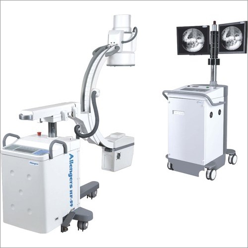
A C-Arm Machine is a medical imaging device widely used in hospitals and diagnostic centers to provide real-time X-ray images (fluoroscopy) during surgeries, diagnostics, and interventional procedures.
🔍 What is a C-Arm?
A C-Arm is named after its C-shaped arm that connects the X-ray source and detector. This design allows flexible movement around the patient to capture images from different angles without repositioning the patient.
⚙️ Main Components
- X-ray Generator – Produces X-rays
- Image Intensifier / Flat Panel Detector – Converts X-rays into images
- C-shaped Arm – Enables multi-angle imaging
- Monitor Display System – Shows real-time images
- Control Panel – Adjusts exposure, angle, and imaging settings
🏥 Applications
C-Arm machines are commonly used in:
- Orthopedic surgeries (fractures, implants, joint procedures)
- Cardiology (angiography, stent placement)
- Pain management procedures
- Urology & Gastroenterology
- Neurosurgery
🔄 Types of C-Arm Machines
- Mobile C-Arm
- Portable and easy to move
- Used in operation theatres
- Most common in small to mid-size hospitals
- Fixed C-Arm
- Installed permanently in cath labs
- Used for advanced cardiac procedures
📊 Key Features
- Real-time fluoroscopy imaging
- High-resolution digital images
- Low radiation exposure (with modern systems)
- 360° rotation for better positioning
- Image storage & processing
⚡ Advantages
- Improves surgical accuracy
- Reduces operation time
- Minimally invasive procedures
- Better patient outcomes
💰 Approx Price (India)
- Mobile C-Arm: ₹12 lakh – ₹60 lakh
- Fixed C-Arm: ₹1.5 crore – ₹5 crore+
📌 Quick Tip for Your Business (Parbhriti Medical Systems)
If you’re selling or promoting C-Arm machines, highlight:
- Low radiation dose
- High image clarity
- Easy mobility
- After-sales service
II C-ARM
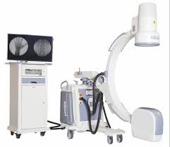
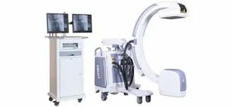
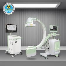
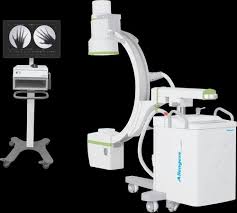
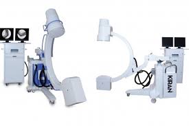
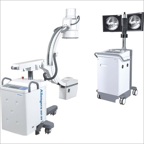
An II C-Arm Machine (Image Intensifier C-Arm) is a mobile X-ray imaging system widely used in hospitals for real-time imaging (fluoroscopy) during surgeries and diagnostic procedures.
🔷 What does “II” mean?
II = Image Intensifier
It is the component that converts low-dose X-rays into a bright visible image, allowing doctors to see live internal structures.
🔷 Main Parts of II C-Arm Machine
- C-shaped arm
- Holds X-ray tube and image intensifier
- Moves in multiple directions (AP, lateral, oblique)
- X-ray Tube
- Generates X-rays
- Image Intensifier (II)
- Converts X-rays → visible light → digital image
- Monitor System
- Displays real-time images
- Control Panel
- Adjusts exposure (kV, mA), fluoroscopy mode, etc.
🔷 Working Principle
- X-rays pass through the patient
- Image Intensifier captures transmitted X-rays
- Converts them into bright image
- Image displayed on monitor in real-time
🔷 Applications
- Orthopedic surgeries (fractures, implants)
- Pain management procedures
- Cardiology
- Urology
- Gastroenterology
- Vascular surgeries
🔷 Types of II C-Arm
- 6 inch / 9 inch / 12 inch Image Intensifier
- Mobile C-Arm
- Fixed C-Arm (OT-based)
🔷 Key Features
- Real-time fluoroscopy
- Low radiation dose (compared to conventional)
- Digital image storage
- Easy mobility
- Multi-angle positioning
🔷 Advantages
✔ Live imaging during surgery
✔ Faster procedures
✔ Better accuracy
✔ Reduced surgical risk
🔷 Limitations
❌ Lower image quality compared to Flat Panel DR C-Arm
❌ Image distortion at edges
❌ Bulky Image Intensifier
🔷 II vs Flat Panel C-Arm
| Feature | II C-Arm | Flat Panel C-Arm |
|---|---|---|
| Technology | Image Intensifier | Flat Panel Detector |
| Image Quality | Good | Excellent |
| Distortion | Present | Minimal |
| Cost | Lower | Higher |
FPD C-ARM
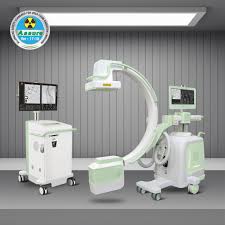
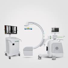
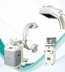
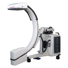
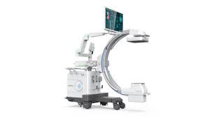
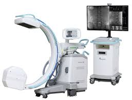
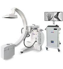
An FPD C-Arm machine is an advanced type of C-arm X-ray system that uses a Flat Panel Detector (FPD) instead of a traditional image intensifier.
🔍 What is an FPD C-Arm Machine?
An FPD (Flat Panel Detector) C-arm is a mobile fluoroscopy imaging system used in operation theatres for real-time X-ray imaging during surgeries like orthopedics, urology, pain management, etc.
👉 The “C-arm” shape connects:
- X-ray tube (one side)
- Detector (other side)
⚙️ Key Features of FPD C-Arm
- Flat Panel Detector (FPD):
Provides high-resolution, distortion-free digital images - Better image quality:
High dynamic range & low noise for clear anatomy visualization - Low radiation dose:
Advanced technology reduces patient exposure - Real-time imaging (Fluoroscopy):
Helps surgeons during procedures - Large Field of View:
Example: 9″x9″ or larger detectors for full anatomy coverage - Digital storage & processing:
Images can be stored, zoomed, and enhanced
🏥 Applications
Used in:
- Orthopedic surgeries (fracture fixation, implants)
- Urology procedures
- Gastroenterology
- Pain management
- Vascular & general surgeries
⚡ Difference: FPD vs Image Intensifier C-Arm
| Feature | FPD C-Arm | Image Intensifier C-Arm |
|---|---|---|
| Image Quality | Very high (HD digital) | Moderate |
| Distortion | No distortion | Possible distortion |
| Radiation Dose | Lower | Higher |
| Size | Compact & modern | Bulky |
| Maintenance | Low | Higher |